In-Office Procedures
Click on a test below to learn more
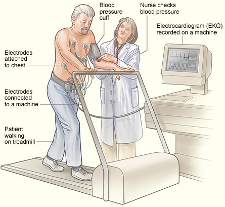
An Exercise Stress Test records the heart’s electrical activity (rate and rhythm) during exercise.
Electrodes will be placed on the chest similar as for an electrocardiogram (EKG). Your blood pressure, heart rate, and EKG will be recorded at rest, usually while you are lying on your back and again when standing.
You will then be asked to perform an exercise test on a motor-driven treadmill. The exercise-protocol that you will follow will be determined by the cardiologist supervising your test, but will begin at a relatively easy level and become progressively more difficult with each subsequent stage.
Your blood pressure, heart rate, and EKG will be recorded at frequent intervals during exercise and after exercise. The physicians or technologist may stop the test at any time for medical reasons.
You may ask to stop the test at any time because of significant fatigue or discomfort. However, we encourage you to exercise as long as possible so that we may assess your heart under maximum exercise stress.
All patients are required to return to the office for their results. Dr. Husain and Patti Cox, N.P. will discuss the results in detail with the help of diagrams and charts. All questions will be answered and the need for further testing or adjustment of medications will be addressed. For the safety and privacy of our patients, results cannot be given or explained over the phone.
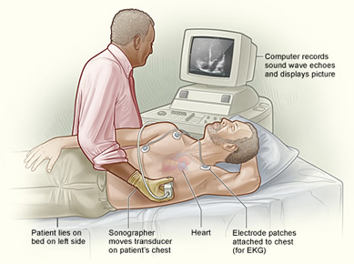
Stress echocardiography is a test that uses ultrasound imaging to show how well your heart muscle is working to pump blood to your body.
A resting echocardiogram will be done first. While you lie on your left side with your left arm out, a small device called a transducer is held against your chest. A special gel is used to help the ultrasound waves get to your heart.
After completion of the echocardiogram you will be asked to walk on a treadmill. Slowly (about every 3 minutes), you will be asked to walk faster and on an incline. In most cases, you will need to walk for around 5 to 15 minutes, depending on your level of fitness and your age. Your doctor will ask you to stop:
As soon as the treadmill is stopped, you will be asked to quickly get back on the exam table and the echocardiogram you had at the beginning of the test will be quickly repeated.
The test usually takes 30-45 minutes.
All patients are required to return to the office for their results. Dr. Husain and Patti Cox, N.P. will discuss the results in detail with the help of diagrams and charts. All questions will be answered and the need for further testing or adjustment of medications will be addressed. For the safety and privacy of our patients, results cannot be given or explained over the phone.
A chemical stress test is obtained for patients who cannot walk on a treadmill. The chemical most frequently used is called Lexiscan. At first, a radionucleatide tracer (usually Cardiolite) is utilized. Images are obtained before the Lexiscan is infused and after the Lexiscan is infused. This helps to determine if areas of the heart are receiving enough blood or if there are blockages (coronary artery disease).
The patient will receive an IV to allow the injection of the tracer. There will be a wait of approximately an hour to an hour and a half before the first images are taken. Patients will then have electrodes (patches) placed on the chest. Several EKGs will be obtained. The patient will receive the medication Lexiscan through the IV, replacing the exercise portion of the test. The Lexiscan is injected into the IV. This is followed by an injection of the tracer via of the IV. Images will be obtained an hour and a half after the Cardiolite injection. The medication Lexiscan will dilate the heart’s arteries. If the heart’s arteries are healthy they will dilate more than arteries that are not healthy. The patient may feel flushed, chest pressure or pain, shortness of breath, and or headache.
It is important to follow the preparation of the Lexiscan/Cardiolite stress test. For 24 hours before the test the following cannot be taken: coffee, tea, colas, chocolates or candies, frosting, cookies, pies, cocoa, milk that contains chocolate, aspirin products that contain caffeine such as Anacin or Excedrin, persantine (dipyridamole), theophylline or theophylline containing products such as Constant-T, Primatene, Quibron, Slo-phylline, Theo-dur and aggrenox. Drinks cannot be taken if they are labeled “ caffeine free” or “decaffeinated”. It is important not to have any caffeine 24 hours before the test. The patient may not eat or drink after midnight. 4-6 hours should be allowed for this test.
All patients are required to return to the office for their results. Dr. Husain and Patti Cox, N.P. will discuss the results in detail with the help of diagrams and charts. All questions will be answered and the need for further testing or adjustment of medications will be addressed. For the safety and privacy of our patients, results cannot be given or explained over the phone.
The echocardiogram is a non-invasive, painless procedure which uses a transducer to transmit high-frequency sound waves (ultrasound) within the heart chambers and valves. These waves bounce throughout the heart area and produce the images that detect heart damage and/or disease.
The primary use of an echocardiogram is to measure structure and function of the heart.
Clothing from the upper body is removed and covered by a gown or sheet to keep you comfortable and maintain the privacy of females.
The patient then lies on an examination table or a hospital bed.
Sticky patches or electrodes are attached to the chest and shoulders and connected to electrodes or wires.
A colorless gel is then applied to the chest and the echo transducer is placed on top of it.
The echo technologist then makes recordings from different parts of the chest to obtain several views of the heart.
You may be asked to move from your back and to the side. Instructions may also be given for you to breathe slowly or to hold your breath. This helps in obtaining higher quality pictures. The images are constantly viewed on the monitor.
The test usually takes 15-20 minutes.
All patients are required to return to the office for their results. Dr. Husain and Patti Cox, N.P. will discuss the results in detail with the help of diagrams and charts. All questions will be answered and the need for further testing or adjustment of medications will be addressed. For the safety and privacy of our patients, results cannot be given or explained over the phone.
An arterial duplex ultrasound uses sound waves to create a color map of the arteries in your leg(s) to identify narrowing of your vessels that may be causing leg pain when walking, resting leg pain, buttock/hip pain, foot, ankle, heel or toe ulcers, or skin discoloration.
This test will show the arteries in your legs, evaluating the size and shape of arteries, blood flow through the arteries and any plaque or obstructions within the arteries that may create blood flow blockages.
The test is performed using a hand-held probe and skin gel, which passes over the entire leg down to the ankle, creating an image. You will be asked to lie flat on your back for 20-30 minutes.
All patients are required to return to the office for their results. Dr. Husain and Patti Cox, N.P. will discuss the results in detail with the help of diagrams and charts. All questions will be answered and the need for further testing or adjustment of medications will be addressed. For the safety and privacy of our patients, results cannot be given or explained over the phone.
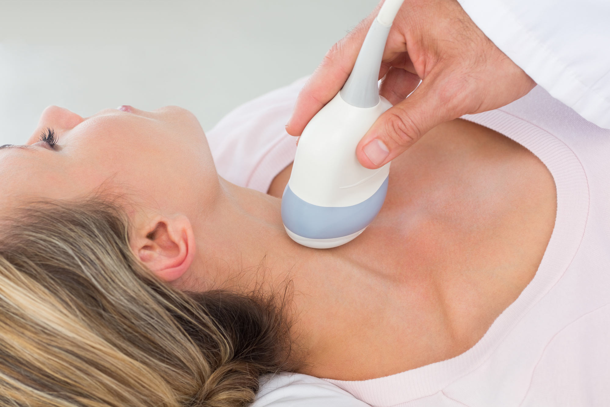
Carotid (ka-ROT-id) ultrasound is a painless and harmless test that uses high-frequency sound waves to create pictures of the insides of your neck arteries.
This test will evaluate the size of the arteries, blood flow through the arteries and any plaque or obstructions within the arteries that may create blood flow blockages.
The test is performed using a hand-held probe and skin gel, which passes over the base of the neck, just below the ear, creating an image.
You will be asked to lie flat on your back for 10-20 minutes.
All patients are required to return to the office for their results. Dr. Husain and Patti Cox, N.P. will discuss the results in detail with the help of diagrams and charts. All questions will be answered and the need for further testing or adjustment of medications will be addressed. For the safety and privacy of our patients, results cannot be given or explained over the phone.
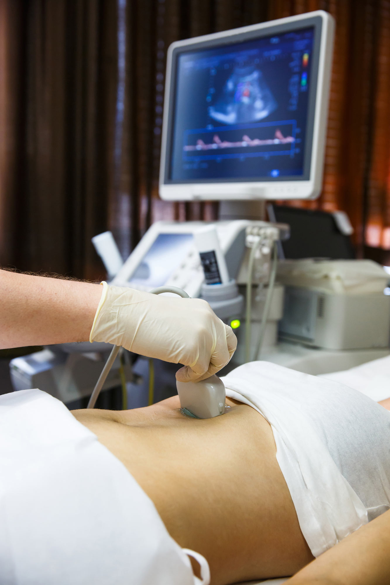
An abdominal vascular ultrasound is a test that evaluates the aorta, the largest artery in the abdomen as well as its branches.
The test will help evaluate the size and blood flow of the aorta and its branches. It will help look for an enlargement (aneurysm), look for blockages or narrowing and help monitor existing aneurysms or arterial diseases as well as follow up after surgical treatment.
You will be asked to lie down on an examination table. The technician will place a clear gel on your abdomen. The gel allows the transducer (a device that both puts out and detects ultrasound signals) to slide around easily on your skin. When the transducer is placed against the skin, an image of the artery is shown on a video screen. The exam usually takes 20-30 minutes to complete.
All patients are required to return to the office for their results. Dr. Husain and Patti Cox, N.P. will discuss the results in detail with the help of diagrams and charts. All questions will be answered and the need for further testing or adjustment of medications will be addressed. For the safety and privacy of our patients, results cannot be given or explained over the phone.
Renal artery ultrasound is a test that shows the renal arteries, the arteries that carry blood to the kidney. These arteries may narrow or become blocked and this may result in kidney failure or high blood pressure (hypertension). Ultrasound waves are used to make an image of the artery.
This is a diagnostic test that produces images of kidney. This test will evaluate the size and flow of blood in the arteries, tissues and any obstructions within the arteries that may create blood flow blockages.
You will be asked to lie down on an examination table. The technician will place a clear gel on your abdomen. The gel allows the transducer (a device that both puts out and detects ultrasound signals) to slide around easily on your skin. When the transducer is placed against the skin, an image of the artery is shown on a video screen. The exam usually takes 20-30 minutes to complete.
All patients are required to return to the office for their results. Dr. Husain and Patti Cox, N.P. will discuss the results in detail with the help of diagrams and charts. All questions will be answered and the need for further testing or adjustment of medications will be addressed. For the safety and privacy of our patients, results cannot be given or explained over the phone.
They are small electronic devices that monitor and record the electrical activity in your heart through electrodes that are placed on your body and will help your Cardiologist in diagnosing a potential heart rhythm abnormality.
A Holter monitor is for short-term monitoring, usually 24-48 hours.
An Event monitor or an MCOT monitor is used for longer-term monitoring, usually from 1-4 weeks.
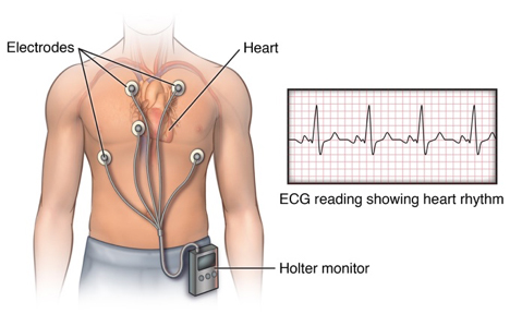
While the cardiac monitor is attached, you will go about your regular activities, and return the device to your Cardiology office at the end of the procedure. The data taken from the monitor about when arrhythmias occur and where in the heart they are coming from can help your Cardiologist diagnose and treat any problems.
While a patient is wearing a monitor here are a few important details to keep in mind to ensure the monitor gets a clear reading:
All patients are required to return to the office for their results. Dr. Husain and Patti Cox, N.P. will discuss the results in detail with the help of diagrams and charts. All questions will be answered and the need for further testing or adjustment of medications will be addressed. For the safety and privacy of our patients, results cannot be given or explained over the phone.
Our office offers complete pacemaker and defibrillator interrogation and reprogramming.
Patients with pacemakers and defibrillators are scheduled for their checks in the office as arranged by Dr. Husain and Patti Cox, N.P.
We also offer in-home monitoring for certain devices.
Dr. Husain or Patti Cox, N.P. will typically see the patients and go over the results of their pacemaker and defibrillator checks. An appointment for the next check will be made at check out.
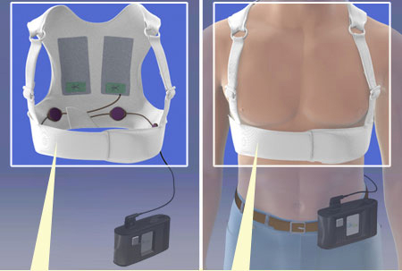
Certain patients are recommended to wear an external defibrillator due to their cardiac condition. Our office is actively involved in identifying those patients, arranging for their installation and providing management of patients who wear an wearable external defibrillator.
Hospital Procedures
Click on a test below to learn more
Cardiac catheterization is a test to check your heart. This test can include a coronary angiogram, which checks the coronary arteries.
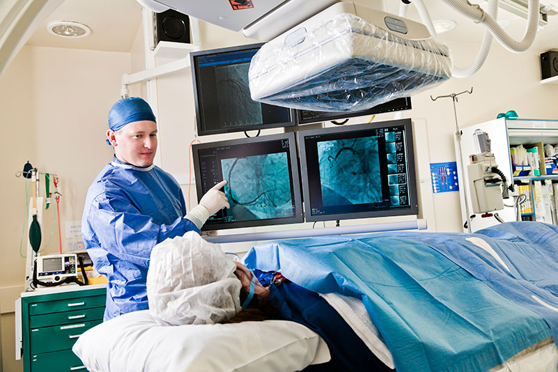
A cardiac catheterization can check blood flow in the coronary arteries, check blood flow and blood pressure in the chamber of the heart, find out how well the heart valves work, and check for defects in the way the wall of the heart moves.
A coronary angiogram is used to find out if you have disease in your coronary arteries (atherosclerosis). If you have atherosclerosis, this test can pinpoint the size and location of fat and calcium deposits (plaque) that are narrowing your coronary arteries.
Results from a coronary angiogram help determine whether treatment with medicines, percutaneous coronary intervention (PCI), such as angioplasty/stenting or coronary artery bypass may be effective.
Angioplasty and related techniques are known as percutaneous coronary intervention (PCI). Angioplasty is a procedure in which a narrowed section of the coronary artery is widened. Angioplasty is less invasive and has a shorter recovery time than bypass surgery, which is also done to increase blood flow to the heart muscle but requires open-chest surgery. Most of the time stents are placed during angioplasty.
An angioplasty is done using a thin, soft tube called a catheter. A doctor inserts the catheter into a blood vessel in the groin or wrist. The doctor carefully guides the catheter through blood vessels until it reaches the blocked portion of the coronary artery.
Cardiac catheterization is done first to identify any blockages.
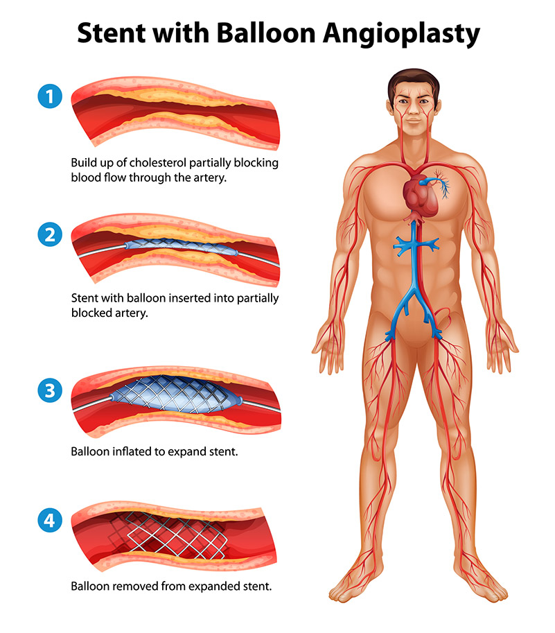
A small, expandable tube called a stent is often permanently inserted into the artery during angioplasty. A very thin guide wire is inside the catheter. The guide wire is used to move a balloon and the stent into the coronary artery. A balloon is placed inside the stent and inflated, which opens the stent and pushes it into place against the artery wall. The balloon is then deflated and removed, leaving the stent in place. Because the stent is mesh-like, the cells lining the blood vessel grow through and around the stent to help secure it.
Stenting should:
Stent placement is standard during most angioplasty procedures. Your doctor may use a bare metal stent or a drug-eluting stent. Drug-eluting stents are coated with medicine that helps keep the artery open after angioplasty.
The procedure may take 30 to 90 minutes. But you need time to get ready for it and time to recover. It can take several hours total. After angioplasty, you will be moved to a recovery room or to the coronary care unit. Your heart rate, pulse, and blood pressure will be closely monitored and the catheter insertion site checked for bleeding. You may have a large bandage or a compression device on your groin or arm at the catheter insertion site to prevent bleeding. You will be instructed to keep your leg straight if the insertion site is near your groin area.
You can mostly likely start walking within 12 to 24 hours after angioplasty. You will likely stay one night in the hospital. You may resume exercise and driving after several days.
You will take antiplatelet medicines to help prevent another heart attack or a stroke. If you get a stent, you will probably take aspirin plus another antiplatelet such as clopidogrel (Plavix). If you get a drug-eluting stent, you will probably take both of these medicines for at least one year. If you get a bare metal stent, you will take both medicines for at least one month but maybe up to one year. Then, you will likely take daily aspirin long-term. If you have a high risk of bleeding, your doctor may shorten the time you take these medicines.
After your procedure, you might attend a cardiac rehabilitation program. In cardiac rehab, a team of health professionals provides education and support to help you recover and build new, healthy habits, such as eating right and getting more exercise. For keeping your heart healthy and your arteries open, making these changes is just as important as getting treatment.
A peripheral angiogram is a test that uses X-rays and dye to help your doctor find narrowed or blocked areas in one or more of the arteries that supply blood to your legs.
Doctors use a peripheral angiogram if they think blood is not flowing well in the arteries leading to your legs or, in rare cases, to your arms. The angiogram helps you and your doctor decide if a surgical procedure is needed to open the blocked arteries.
Peripheral angioplasty is one such procedure. It uses a balloon catheter to open the blocked artery from the inside. A stent, a small wire mesh tube, is generally placed in the artery after angioplasty to help keep it open. Bypass surgery is another procedure. It re-routes blood around the blocked arteries.
Serious risks and complications from peripheral angiograms are very unlikely. But in rare cases:
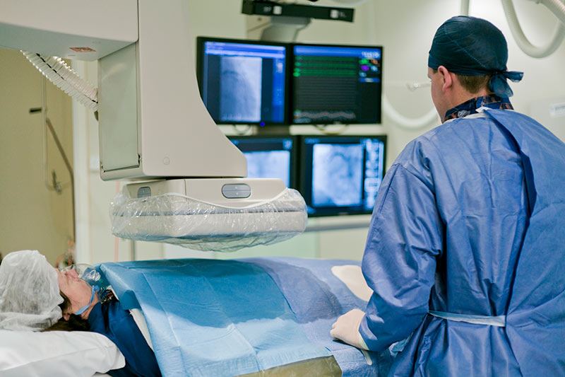
A doctor with special training performs the test with a team of nurses and technicians. The test is performed in a hospital or outpatient clinic.
The TEE test takes a detailed picture of your heart and its major blood vessels. This test helps to detect heart valve disease, heart tumors, and blood clots inside the heart. It also helps detect an aneurysm, which is a swelling, like a balloon, in a blood vessel. TEE stands for trans-esophageal echocardiogram.
The TEE test combines several procedures. It makes a picture by using sound waves that pass through skin and tissue without being heard or felt. This is called ultrasound. The special instrument that sends and receives the sound waves is called a transducer.
A tiny transducer is attached to the end of a flexible tube with a light. The tube is called an endoscope. The scope is passed into your mouth, through your esophagus (food pipe), and down behind your heart. The word “trans-esophageal” means “through the esophagus.”
The transducer sends sound waves into your heart and blood vessels and then receives the signals that bounce back, or “echo.” The signals are converted into pictures that show on a screen. This part of the test is called an echocardiogram.
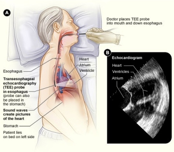
A doctor will perform your TEE test with the help of a nurse or technician. The technician will help you onto a bed and ask you to lie on your left side. You will be connected to machines that monitor your blood pressure, heart rate, and oxygen levels during the test. You will be given a mild sedative by an intravenous line (IV) to help you relax. You’ll remain somewhat alert so you can cooperate with the doctor and staff.
The TEE test may cause some mild discomfort, but it should not be painful. The back of your throat will be numbed with an anesthetic spray. Your throat will feel cool, and you may get a bitter taste in your mouth. You will need to remove any full or partial dentures. To protect your teeth, a plastic mouth guard will be placed in your mouth. You’ll need to press your lips around the guard.
The doctor then will insert the scope into the back of your mouth. You will be asked to swallow the scope, and you may gag for a few moments. The doctor will advance the scope into your esophagus about 12 to 20 inches. Images will be obtained for about half an hour.
You cannot eat or drink for at least 1 hour after the test. The numbing effect of the anesthetic takes this long to wear off. Your nurse will check your gag reflex often by touching the back of your mouth with a tongue depressor. You must be able to gag to keep from choking on food or fluid.
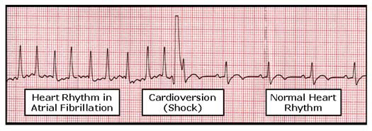
Cardioversion is a corrective procedure where an electrical shock is delivered to the heart to convert, or change, an abnormal heart rhythm back to normal sinus rhythm. Most elective or "non-emergency" cardioversions are performed to treat atrial fibrillation (A Fib) or atrial flutter (AFL), non-life threatening abnormal rhythms in the top of the heart. Cardioversion is also used in emergency situations to correct an abnormal rhythm when it is accompanied by faintness, Low Blood Pressure, chest pain, difficulty breathing, or loss of consciousness.
Each normal heartbeat starts in an area of the heart known as the sinus node, located in the upper right chamber of the heart (right atrium). The sinus node sends organized electrical signals through the heart resulting in a perfectly timed, rhythmic heartbeat. In people with atrial fibrillation however, this electrical signal is chaotic, causing the atria to fibrillate (or "quiver"). This typically results in a fast and irregular heartbeat. While some people have no symptoms, others may experience shortness of breath, lightheadedness and fatigue. Depending on your specific medical history and symptoms, your doctor may recommend a cardioversion to return your heart to normal sinus rhythm.
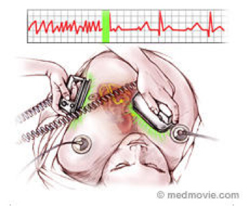
Also known as "direct-current" or DC cardioversion, a synchronized (perfectly timed) electrical shock is delivered through the chest wall to the heart through special electrodes or paddles that are applied to the skin of the chest and back. The goal of the procedure is to disrupt the abnormal electrical circuit(s) in the heart and thereby to reset the heart to normal rhythm. This split second interruption of the abnormal beat allows the heart's electrical system to regain control and restore a normal heartbeat. Electrical cardioversion is performed in a hospital setting where oxygen levels, blood pressure and heart rhythm are closely monitored.
Normal sinus rhythm can be restored more than 90 percent of the time, however atrial fibrillation or other abnormal rhythms may recur in over time. Your doctor may prescribe medications or recommend catheter ablation to reduce the risk of atrial fibrillation recurrence.